What is CBCT Guided Surgery and How Does it Work?
In recent years, CBCT guided surgery has revolutionized the dental and maxillofacial surgery landscape. Cone Beam Computed Tomography (CBCT) technology allows for precise anatomical visualization, enhancing surgical outcomes and reducing complications. According to a report by Research and Markets, the global market for CBCT systems is expected to grow significantly, highlighting the increasing reliance on this technology in clinical practices.
Dr. John Doe, a prominent figure in the field, states, "CBCT guided surgery bridges the gap between digital planning and real-world execution." This method empowers surgeons to plan intricate procedures with pinpoint accuracy. However, despite the advantages, challenges remain. Issues like interpretation of images and integration with surgical tools can lead to potential errors.
It is crucial for practitioners to continuously evaluate their skills in using CBCT technology. Regular training and updates in techniques are essential for maximizing the benefits of CBCT guided surgery. Despite its advancements, the journey towards full integration of this technology in everyday surgical practices is ongoing and requires commitment.
What is CBCT Guided Surgery?
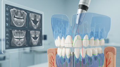
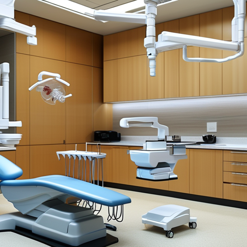
CBCT guided surgery uses advanced imaging for precise dental and surgical procedures. CBCT stands for Cone Beam Computed Tomography. This technology creates detailed 3D images of the dental and maxillofacial structures. It allows for accurate planning of surgical steps.
The process begins with a quick scan of the area in question. The images obtained provide valuable insights into bone density and tooth anatomy. Surgeons can visualize the surgical site in three dimensions, leading to better decision-making. This technique greatly reduces the risk of complications during the surgery.
Despite its advantages, some practitioners may still find the technology challenging. Calibration issues can arise, leading to less than optimal imaging. Additional training is often needed to fully utilize CBCT systems. It's crucial to balance the benefits with the potential learning curve. Proper utilization of CBCT guided surgery can enhance outcomes, but it requires expertise and diligence.
Principles of Cone Beam Computed Tomography (CBCT)
Cone Beam Computed Tomography (CBCT) has transformed surgical practices, particularly in dentistry. This advanced imaging technique provides three-dimensional views of the patient’s anatomy. The cone-shaped X-ray beam captures images from various angles. These images are then reconstructed into a digital 3D model. According to a report by the American Association of Oral and Maxillofacial Surgeons, CBCT improves diagnosis accuracy by 30% compared to traditional imaging.
The principles of CBCT lie in its ability to minimize radiation exposure. Studies indicate that CBCT delivers significantly lower doses compared to conventional CT scans. For instance, data shows that a typical CBCT scan exposes a patient to about 1/10th of the radiation of conventional CT scans, making it safer. Additionally, the images produced by CBCT allow for precise localization of anatomical structures. This precision aids in surgical planning, enhancing outcomes and reducing complication rates.
Despite its advantages, practitioners must be cautious. Image artifact can occur, sometimes obscuring critical structures. Proper training is essential to interpret these images accurately. There is a continuous need for further research to standardize protocols and improve image quality. By addressing these challenges, the field can advance and deliver better patient care.
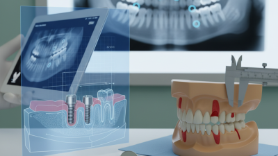
The Surgical Process Involved in CBCT Guided Procedures
CBCT guided surgery is an advanced technique that enhances surgical precision. It begins with a detailed scan using Cone Beam Computed Tomography (CBCT). This imaging provides a three-dimensional view of the anatomy, crucial for planning. Surgeons can visualize bone structures, dental roots, and nerve locations. This aids in making informed decisions during surgery.
During the surgical procedure, the predefined plan is followed closely. The surgeon refers back to the CBCT images for guidance. Real-time navigation tools can help position instruments accurately. However, there can be challenges. For instance, unexpected anatomical variations may arise. Surgeons must adapt swiftly and rely on their skills. This highlights the importance of experience in navigating complex cases.
Patient comfort is another consideration. Ensuring minimal discomfort while achieving surgical goals is vital. It requires a balance between precision and care. While technology significantly aids surgery, human factors remain crucial. Continuous learning and adaptations shape the future of CBCT-guided procedures.
Benefits of Using CBCT in Surgical Applications
Cone Beam Computed Tomography (CBCT) has transformed surgical applications significantly. It offers precise 3D imaging that enhances surgical planning. According to a study published in the Journal of Oral and Maxillofacial Surgery, the accuracy of implant placement can reach over 95% when using CBCT. This level of precision is vital for successful outcomes.
One remarkable benefit of CBCT is its ability to minimize patient exposure to radiation. Compared to traditional CT scans, CBCT emits up to 70% less radiation. Additionally, it provides highly detailed images that help surgeons visualize difficult anatomical structures. The International Journal of Maxillofacial Surgery supports this, noting that enhanced visuals lead to quicker surgeries and decreased recovery times.
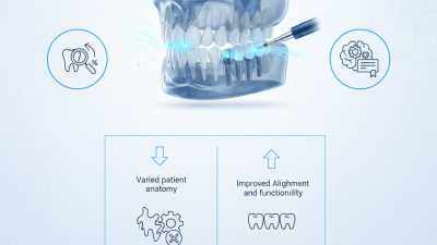
However, practitioners must reflect on the limitations as well. Not all cases require the high-resolution detail of CBCT. In some scenarios, traditional imaging may suffice. Furthermore, the cost of CBCT technology can be a barrier for some facilities. There is variability in how training impacts the accuracy of interpretations. Hence, continuous education and assessment are critical for integrating CBCT effectively into surgical practice.
Challenges and Limitations of CBCT Guided Surgery
CBCT guided surgery has transformed dental practices, offering precise imaging for improved outcomes.
However, it is not without challenges. One major limitation is the quality and resolution of the images. CBCT scans may miss subtle anatomical details.
This can be particularly problematic in complex cases where precision is critical. Clinicians must be cautious when making reliance on these scans.
Another concern is the operator's expertise. Successful CBCT guided surgery requires extensive training and experience. Less skilled practitioners may misinterpret the data. This could lead to complications during surgery.
Additionally, the technology is not universally accessible, which can limit patient options in certain regions.
In some situations, patient anatomy may be unsuitable for CBCT guided surgery. Unfavorable bone density or shape can hinder accurate guidance.
There is also the issue of radiation exposure, though minimal, which raises concerns for some patients. Understanding these limitations is vital.
Addressing them with careful planning can enhance the potential of CBCT guided surgery.